Talipes equinovarus (TEV) is a common, but little known developmental disorder of the lower limb. Get to know about this condition in detail, including its symptoms, causes, diagnosis and treatment options.
Talipes equinovarus Definition
Page Contents
- 1 Talipes equinovarus Definition
- 2 Talipes equinovarus Synonyms
- 3 Talipes equinovarus Incidence
- 4 Talipes equinovarus Types
- 5 Talipes equinovarus Symptoms
- 6 Talipes equinovarus Causes
- 7 Talipes equinovarus Diagnosis
- 8 Talipes equinovarus Treatment
- 9 Talipes equinovarus Prognosis
- 10 Famous people with Talipes equinovarus
It is a congenital anomaly of the foot and ankle. In Latin, talipes refers to “taluspes” meaning ankle and foot and equinovarus suggests an elevated heel turned inwards.
Talipes equinovarus Synonyms
The disorder is also referred to by other names like:
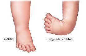
Picture 1 – Talipes equinovarus
- Club foot
- Congenital talipes equinovarus
Talipes equinovarus Incidence
Clubfoot is a common defect present at birth and occurs in every 1,000 live births. Bilateral TEV can be found be found in nearly 50% of cases. About twice as many males are born with the congenital form than females.
Talipes equinovarus Types
The disorder can be differentiated into following two types:
- Postural Talipes equinovarus
- Structural Talipes equinovarus
Talipes equinovarus Symptoms
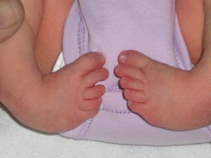
In this condition, the foot of a suffering infant point downwards at the ankle and the heel is turned inwards. The arch of the foot is more pronounced. The middle portion of the foot is also twisted inwards and this makes it look apparently short as well as wide than the other foot. The foot remains fixed in this unusual position and fails to acquire the normal posture, despite efforts, forcing patients to walk on their ankle or toes. In aggressive cases, the foot may appear upside-down. The calf muscles are usually underdeveloped in the affected newborns. There is no sign of discomfort or pain in the foot when the patient attempts to walk. In this condition, deformities usually occur in the following joints of the foot and ankle:
- Subtalar joint
- Talonavicular joint
- Ankle joint
Talipes equinovarus Causes
Each type of TEV has a different etiology. The postural form of the condition is believed to be a consequence of a normal foot held in a deformed position in the uterus. The problem most likely manifests during the final trimester of pregnancy due to frequent contractions within the uterus possibly caused by conditions such as:
Oligohydramnios
It is a condition observed in pregnancy that is characterized by a deficiency of amniotic fluid.
Amniotic band syndrome
A partial rupture of the amniotic sac can cause the fibrous bands of the amnion to encircle and trap the limbs of the fetus only to inhibit its growth, and development.
The cause of structural TEV has been traced to a genetic disorder called Edwards syndrome, in which there are three copies of chromosome 18. Compartment syndrome is another identifiable cause of clubfoot. The causative disorder occurs when increased pressure within a muscle compartment of the fetal leg obstructs the blood from supplying oxygen and nutrients to the compressed tissues. Failure to release the pressure can result in permanent shortening of the ankle joint. Structural deformity of the foot could also be associated with various congenital conditions like:
Breech presentation
The fetus lies longitudinally inside the uterus with the buttocks or feet nearer to the cervix as opposed to the normal head presentation. Most breech babies are born healthy, but there is a slightly elevated risk of a birth defect.
Ehlers Danlos Syndrome
The hypermobility form of the genetic disorder can affect the joint of the foot and dislocate it.
Loeys-Dietz Syndrome
Abnormal organization of blood vessels present as a birth defect can upset the development of the lower extremities in some infants.
Spina bifida
Incomplete formation of the fetal spinal cord can erode the function of the nerves and give rise to a number of orthopedic abnormalities, including club foot.
Talipes equinovarus Diagnosis
Physicians normally look for signs of TEV after the birth of a baby. Further confirmation can be done with an X-ray. Anteroposterior and lateral standing X-rays are normally recommended to know the position of the bones as well as determine any abnormality. Health specialists may often use a grading system called Pirani score to evaluate the severity of the foot deformity. The advanced features of ultrasound scanning during pregnancy enable prenatal diagnosis of clubfoot.
Talipes equinovarus Treatment
The degree of anomaly, associated conditions and secondary muscular changes are the principal factors that must be considered before initiating the treatment of TEV. Healthcare providers usually aim for less use of an invasive procedure in order to avoid complications.
Non-surgical therapy
Ponseti method is a conservative technique used to correct congenital clubfoot without the need of a surgery. In this procedure, an experienced specialist manipulates the patient’s foot into a position in which the deformity is corrected as much as possible. The technique is painless and does not cause any additional discomfort. Once the affected foot is held in the proper position, a plaster cast is used to immobilize it. The traditional cast needs to be changed every 1-3 weeks until the child’s foot moves to a normal position. Foot position drastically improves with the help of repeated manipulation and plaster casting. Achilles tenotomy, a minor surgery, would be necessary as a part of Ponseti regime to release the tight Achilles tendon at the back of the foot. The tendon is stretched and the heel gradually drops down. Orthoses or corrective shoes can be used in case a patient has difficulty walking, and also to prevent TEV from recurring. Another method that can be utilized to normalize the foot deformity is Botox treatment. The chemical is injected into the patient’s calf muscle to help loosen the tight Achilles tendon. Within a span of 4-6 weeks, the foot regains its usual position. However, a Botox injection has only a temporary effect.
Surgical treatment
The conventional form of treatment usually fails to correct the twisted hind foot. The tendons, ligaments and joints in the foot require surgical correction. Complete soft tissue release is normally performed between 6 and 12 months. The anterior tibialis tendon located on the front of the ankle gets inflamed in clubfoot. Centralization of this tendon can readily repair the deformed foot. The other operative methods that would be required later in childhood are:
- Talectomy
- Fusion of the midtarsal and subtalar joints
- Wedge excision of the calcaneocuboid bone
- Calcaneal osteotomy
Talipes equinovarus Prognosis
Approximately 50-60% of neonates with clubfoot show remarkable improvement. However, a small percentage of children may have to opt for a major surgery, which is generally successful. Modern techniques of casting have high success rates even in older children.
Famous people with Talipes equinovarus
The names of some well-renowned people who had been born with this congenital disorder are given below:
Picture 2 – Talipes equinovarus Image
- Thaddeus Stevens
- Damon Wayans
- Gary Burghoff
- Steven Gerrard
- Miguel Riffo
- Matt Lloyd
- Ben Greenberg
- Jennifer Lynch
- George Gordon
Each case of Talipes equinovarus is different and should be dealt with carefully. In the absence of treatment, serious functional and developmental problems can arise in a pediatric patient. To ensure that your child resumes a normal life, immediately get in touch with a doctor for prompt correction of the twisted foot.
References:
http://en.wikipedia.org/wiki/Club_foot
http://www.ncbi.nlm.nih.gov/pmc/articles/PMC1571059/
http://www.medicalnewstoday.com/articles/183991.php
http://www.webmd.com/a-to-z-guides/clubfoot-11038


