Radiocarpal Joints are involved in a lot of flexion and extension activities in the wrist. Know all about this joint, including its anatomy and mechanism.
What is Radiocarpal Joint?
Page Contents
It is the anatomical term used to refer the point of attachment between the carpal bones of the hand and radial bones located in the forearm. This joint is also commonly referred to as the “Wrist Joint”.
Radiocarpal Joint Location
It is a joint located between the proximal carpal bone rows and the distal extremity of the radius.
Radiocarpal Joint Anatomy
The joint is classified as a Synovial one. It is bound together by ligaments and contains a fluid and cartilaginous cavity within the bones that is known to be the synovial capsule. The radiocarpal joint may perform motions that include abduction, adduction, extension and flexion. The hand is tilted upon the wrist from one side to another as well as bent upon the wrist from the front to the back.
The radiocarpal joint consists of four bones in total. These involve:
- Radius
- Scaphoid
- Lunate
- Triquetrum
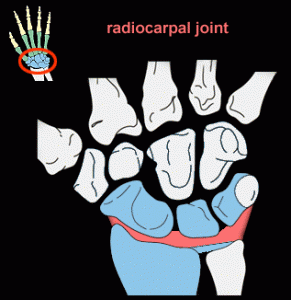
The radius is the extended bone of the forearm the lower or distal end meets the carpal bones of the hand. The Triquetrum, Lunate and the Scaphoid bone cluster together to form the proximal row of the carpus or the bunch of small eight bones located underneath the wrist. The lunate and scaphoid bones meet the radius bone situated in the radiocarpal joint. The Triquetrum does so only once, when the hand is drawn towards the body or bent in the way of the pinky finger. This joint within the carpal and the radial bones is known as an ellipsoid or condyloid joint. This refers to the concave radius surface of the curves surrounding the neighboring convex carpus surface.
Picture 1 – Radiocarpal Joint
Source – classes.kumc.edu
Constituents of the radiocarpal joint may be separated as extrinsic or intrinsic to the joint. A fluid-filled capsule encircled by the synovial membrane is intrinsic to the joint. A gap between the carpus and the radius contains the synovial membrane and runs constant with cavities that are similar and located between and among the carpal bones. This membrane releases a substance known as the synovial fluid that lubricates and fills the joints. The joint cartilage is also situated inside the membrane and acts as a cushion for the space to ensure that there is no friction between the bones. Blood vessels further permeate this space to supply the joint with nutrients.
The wrist ligaments are found outside the radiocarpal joint. Ligaments are mainly composed of collagen or firm connective tissue fibers that connect the bones and shield and encircle the joint. The palmar radiocarpal ligaments can be found in the wrist, on the side of the palm. They run between
- Scaphoid and the Radius
- Lunate and the Radius
- Triquetrum and the Radius
Similarly, the dorsal ligaments on the back of the wrist connect the opposite sides of these bones to the radius. A large articular disk also lies external to the radiocarpal joint and is immediately beside the joint on the pinky-finger or the medial slope of the wrist. It can be found between the Pisiform and Triquetrum bones of the Carpus and the lower or distal end of the ulna bone situated in the forearm.
Radiocarpal Joint Movements
The radiocarpal joint allows multiple wrist motions by connecting the forearm with the hand. Muscles located on the palm side in the anterior forearm can help curl or flex the hand. Muscles positioned on the dorsal side of the posterior forearm assist in extending the hand or twisting it backward. Extra muscles found in the forearm can help abduct or adduct the hand over the wrist, thus shifting it in the way of the pinky or thumb. The simultaneous movement of the radiocarpal joint, the intercarpal joints and the radioulnar joint of the hand can assist in performing more complex motions.
Four major movements are performed with the aid of this joint. Radiocarpal Joint Motions include:
- Extension
- Abduction (radial deviation)
- Adduction (ulnar deviation)
- Flexion
These four movements produce Circumduction taking place in succession. Some of the extension and flexion movements are always attended by similar motion at the Midcarpal Joint. The four motions are performed by muscle group combinations. Flexion is primarily produced by flexor carpi ulnaris and flexor carpi radialis assisted by abductor pollicis longus, palmaris longus and the thumb and finger flexors. The ulnar extensors and the radial extensors aided by the extensors of thumb and fingers produce Extension. The Abductor Pollicis Longus produce abduction. Two radial extensors and the carpi radialis flexor act together to produce abduction when the wrist moves from the midline.
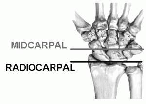
Some of the extension and flexion movement is constantly attended by similar motion at the Midcarpal joint. When flexion occurs, a greater proportion occurs in the Midcarpal joint. A great part of extension happens at the joint of the wrist itself.
The hand, in relation to the forearm, is capable of three kinds of motion. These include
- Flexion and Extension
- Pronation and Supination
- Radial or Ulnar Deviation
The wrist joint has a complicated ligament configuration that helps it maintain mobility without loss of stability.
Radiocarpal Joint Pictures
Want to know how this bone joint looks like? Here are some useful Radiocarpal Joint images that you will find useful for reference. Take a look at these Radiocarpal Joint photos to get an idea about the appearance of this wrist joint.
Picture 2 – Radiocarpal Joint Image
Source – moon.ouhsc.edu
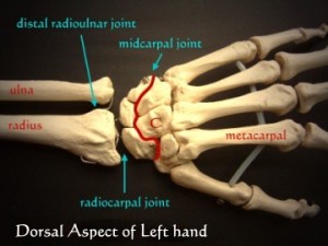
Picture 3 – Radiocarpal Joint Photo
Source – pt.ntu.edu.tw
References:
http://www.mananatomy.com/body-systems/skeletal-system/wrist-joint
http://www.wisegeek.com/what-is-the-radiocarpal-joint.htm
http://www.wisegeek.com/what-is-the-radiocarpal-joint.htm
http://medical-dictionary.thefreedictionary.com/wrist+joint


