What is Ewing Sarcoma?
Page Contents
- 1 What is Ewing Sarcoma?
- 2 Ewing Sarcoma Stages
- 3 Ewing Sarcoma Causes
- 4 Ewing Sarcoma and Genetics
- 5 Ewing Sarcoma Symptoms
- 6 Ewing Sarcoma Prevention
- 7 Ewing Sarcoma Diagnosis
- 8 Ewing Sarcoma Differential Diagnosis
- 9 Ewing Sarcoma Treatment and Management
- 10 Ewing Sarcoma Alternative Treatment
- 11 Ewing Sarcoma Prognosis
- 12 Ewing Sarcoma Incidence
- 13 Ewing Sarcoma Survival Rate
- 14 Ewing Sarcoma Support Groups
Ewing Sarcoma, or Ewing’s Sarcoma, is a type of primary bone cancer that is commonly seen in children and teenagers. The cancerous cells are mainly found in the bones and soft tissues. The tumors generally occur in the femur, the pelvis, the humerus, the clavicle and the ribs.
It is included in the cancer group named Ewing Sarcoma Family of Tumors (ESFT) along with primitive neuroectodermal tumors because both these conditions are usually associated with a common genetic locus. Otherwise, the two diseases are completely different.
This disorder has been named after American pathologist James Ewing who was the first person to describe the tumor. He established it as a separate condition from other cancers like lymphoma.
Ewing Sarcoma Stages
The condition has two primary stages:
Localized tumor
Ewing’s Sarcoma is considered to be a localized condition when it involves only the nearby tissues like the muscles and tendons. The condition is considered to be localized only if the diagnostic studies do not detect the tumor to affect distant organs.
Metastatic tumor
The metastatic stage denotes that the tumor has already spread to various other organs and body parts from its original location. In these cases, the tumor can affect other bones and their bone marrow as well as organs like the lungs, liver, kidney and lymph nodes.
Ewing Sarcoma Causes
The causes behind the development of this cancer are not clearly known. Like many other forms of tumors, it is known to result mainly due to the abnormal growth of certain stem cells which gradually forms the tumor.
Ewing Sarcoma and Genetics
Researchers have shown chromosomal changes in the DNA of a cell to be a possible cause of the disorder. These changes develop in a child after birth instead of being inherited. But the reasons for these changes are not evident.
Certain cells may become cancerous due to genetic exchange between some chromosomes. Translocation between the chromosome 11 and chromosome 12 causes most (85%) of the Ewing’s Sarcoma cases. The EWS gene located on chromosome 22 gets fused with the FLI1 gene located on chromosome 11 due to this translocation.
Ewing Sarcoma Symptoms
The condition can lead to various signs and symptoms depending on the size and location of the tumor. The common symptoms include:
- Pain around the area of the tumor
- Redness and/or swelling around the area of tumor
- Decreased appetite
- Fever
- Weight loss
- Fatigue
- Loss of bladder and bowel control (in cases where the tumor is located in the spine)
- Symptoms associated with nerve compression due to the tumor (including tingling, numbness and paralysis)
A growing tumor can put extreme pressure on the bones and soft tissues around it. This considerably weakens the bones, increasing the risk of bone fractures in a patient.
Ewing Sarcoma Prevention
It is not possible to prevent the disorder because of its unclear etiology. The risk factors responsible for triggering the genetic changes are not known to be associated with any environmental elements or even radiation exposure.
Ewing Sarcoma Diagnosis
Pain and swelling in the legs, arms, back, chest and trunk are the initial symptoms. The condition often goes undiagnosed until it leads to more serious symptoms. In many cases, the disorder is first suspected after the bone weakness becomes evident as the bones break easily after small injuries.
Imaging Tests
Various imaging studies are used for diagnosing this disorder. During these tests, the patients may be given contrast dye injections or asked to drink contrast solutions to develop clear images. These imaging tests include:
X-ray
X-ray images are useful for observing the internal bone structures to detect any abnormality around them. It is very useful for diagnosing the tumors.
CT (Computerized Tomography) scan
A CT scan helps to create cross-sectional images of the bones and soft tissues by combining the x-ray findings. This helps to detect the tumors.
MRI (Magnetic resonance imaging)
This diagnostic procedure is used for producing detailed images of the bones in the affected area. The MRI can help the doctor to determine the stage of the tumor as well as the possibility of nerve or blood vessel involvement and allow him or her to make a treatment plan.
Bone Scan
This test detects bone injury, bone loss and infections by using a harmless amount of radiation. It helps to confirm the diagnosis made by x-ray. Bone scan also helps to determine the metastasis stage of the cancer throughout the skeleton.
PET (Positron Emission Tomography) scan
PET scan also uses radioactive substances for imaging the changes in various cells in the body. The diagnostician injects some radioactive-tagged glucose into the bloodstream to see if any tissue uses more than the normal amount of glucose. These tissues are then scanned for tumors.
Radiographs
Radiographic images can show an “onion skin” appearance of the bones around the tumor due to periosteal reaction.
Biopsy
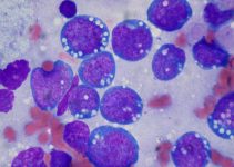
A small sample can be removed from the suspected tumor and sent for examination (biopsy). The sample is then analyzed under a microscope for detecting any cancer cells present. In some cases, a bone marrow biopsy is required to determine whether the cancer has metastasized to bone marrow.
Bone Marrow Aspiration
In this procedure, a diagnostician collects a small quantity of the bone marrow fluid from the hip bones of a patient. Sometimes, solid tissues from the bone marrow may also be collected. The samples are then sent to be tested for determining the size, maturity and number of blood cells as well as the abnormal cells.
Blood Test
A CBC or complete blood count test shows if there are any abnormalities in the blood. Any abnormality in the CBC result may suggest metastasis of the cancer to the bone marrow of a patient. A blood test named Lactate Dehydrogenase or LDH can determine the enzyme levels in the blood. Elevated serum LDH level is associated with the condition. Ewing’s sarcoma may also increase the red blood cell (RBC) sedimentation rate.
Ewing Sarcoma Differential Diagnosis
A diagnostician should take care to rule out the presence of the following disorders, that give rise to similar symptoms, while diagnosing this type of bone cancer:
- Osteosarcoma
- Non-Hodgkin Lymphoma
- Neuroblastoma
- Rhabdomyosarcoma
- Osteomyelitis
- Nonrhabdomyosarcoma Soft Tissue Sarcomas
- Rickets
Ewing Sarcoma Treatment and Management
The treatment of this disorder may vary from one patient to another depending on the stage of severity and present symptoms. The cure often includes various approaches including chemotherapy, surgery and radiation:
Radiation Therapy
It is a painless process similar to x-rays. This therapy uses a machine that kills the cancerous cells by high-energy X-ray beams. The beams often damages some normal cells as well, but the healthy cells in the area can easily repair the damaged ones. The main object of the treatment is to damage the tumor cells beyond any recovery. Radiation therapy is often combined with chemotherapy and surgery (rarely). At present, it uses external radiation for destroying damaged cells. Scientists are carrying out studies to determine whether this treatment can be more effective if the radiation is implanted within the body of patients.
Chemotherapy
This treatment involves the use of various drugs that kills the cancer cells. The drugs can be administered as oral tablets or injected into a muscle or vein. Chemo is referred to as a systemic treatment as the drugs used in the therapy travels to the affected body parts, with the bloodstream, to destroy all the tumor cells throughout the body. It is called combination chemotherapy when it uses multiple medicines. In the treatment of ESFT, chemotherapy is sometimes used to kill the remaining cancer cells after radiation therapy or surgery.
Surgery
Surgical intervention may be required in some cases for removing the tumor. Sometimes, a surgery is performed for removing remnants of the tumors after chemotherapy. Surgery is performed only if it is impossible to clear the tumor by the other treatment options.
Amputation may be necessary in severe cases where the tumor attaches itself to blood vessels and nerves. In this method, the affected limb is surgically removed along with the tumor.
Myeloablative Therapy
Myeloablative Therapy, along with stem cell support, is mainly used as supplement therapy to the treatments mentioned above. It is used only to treat patients with resistant or recurring Ewing’s Sarcoma or a widely disseminated cancer. All rapidly dividing cells in the body are destroyed by this intense chemotherapy regimen. These include hair cells, blood cells as well as the tumor cells. The self-renewing stem cells create all types of blood cells and other important cells that circulate with blood. This treatment involves stem cell support which helps the stem cells to produce these cells in large amounts after the chemotherapy kills all the cancerous cells.
Treatment may include high-dose chemotherapy and stem cell transplantation, depending on the stage and severity of the cancer.
Ewing Sarcoma Alternative Treatment
Various natural and alternative methods, including the following, are used for its treatment:
- Acupuncture
- Massage therapy
- Herbal products
- Vitamins and other nutritional supplements
- Special diets
Ewing Sarcoma Prognosis
The prognosis is determined by a number of factors including the extent, location and size of the tumor, how it responds to therapy as well as the age and health of patients. His or her tolerance of specific procedures, medications and therapies along with the metastasis stage of the cancer also play important roles in determining the outcome.
Early diagnosis and prompt treatment are very important for having the best possible outcome. Patients should receive regular follow-up care for any signs of recurrence.
Ewing Sarcoma Incidence
Three out of every one million people develop the condition every year in the United States. It is most common in individuals aged between 10 and 20 years with the average age of onset being 15 years. However, it can also occur in adults above 30 years of age. The tumor affects both males and females at the ratio of 1.6:1.
Ewing Sarcoma Survival Rate
The survival rate for patients of this disease has increased considerably over time. At present, the survival rate for children aged below fifteen years ranges between 70% and 75%, while patients who are 15 years to 19 years old have a survival rate of 20% to 49%. Individuals who do not develop metastasis have better chances of surviving compared to those with metastasized tumors.
Ewing Sarcoma Support Groups
There are numerous forums and foundations that provide guidelines about this cancer to spread awareness and to help the patients. These organizations include:
American Cancer Society, Inc.
1599 Clifton Road NE
Atlanta, Georgia 30329
United States
Tel: (404)320-3333, (800)227-2345
Website: http://www.cancer.org
National Coalition for Cancer Survivorship
1010 Wayne Avenue
7th Floor
Silver Spring, Maryland 20910
Tel: (301)650-9127
Fax: (301)565-9670
Email: [email protected]
Website: http:// www.canceradvocacy.org
Sarcoma Foundation of America
9884 Main Street
Damascus, MD 20872
USA
Tel: (301)253-8687
Fax: (301)253-8690
Email: [email protected]
Website: http://www.curesarcoma.org
Ewing’s Sarcoma is a life threatening disorder, associated with a considerable mortality rate. Early diagnosis, along with immediate treatment, is required to extend the life expectancy of sufferers. Patients are allowed to return to their normal lifestyle after treatment. However, regular follow-up is necessary to prevent the tumors from recurring.
References:
http://www.
http://www.mayoclinic.org/
http://www.childrenshospital.
http://www.royalmarsden.nhs.
http://orthoinfo.aaos.org/

