Palatine Bone is a small bone found in many animal species. It is an important part of the scull. Read on to know all about it, including its location and function.
What is Palatine Bone?
Page Contents
- 1 What is Palatine Bone?
- 2 Palatine Bone Location
- 3 Palatine Bone Ossification
- 4 Palatine Bone Description
- 5 Palatine Bone Anatomy
- 6 Palatine Bone Articulations
- 7 Palatine Bone in Other Animals
- 8 Palatine Bone Functions
- 9 Horizontal Plate of Palatine Bone
- 10 Perpendicular Plate of Palatine Bone
- 11 Palatine Bone and Cleft Palate
- 12 Palatine Bone Pictures
Various animals belonging to the Chordata phylum have two L-shaped bones that constitute parts of their orbits, hard palate and nasal cavities. Each of these bones is known as a Palatine Bone, commonly referred to as Palatum. It is one of the many paired bones in the human body.
The Palatine bone can be affected by infection, facial fractures, trauma and cancer. Various birth defects can also affect this bone and cause a defect in the face of an individual.
Palatine Bone Location
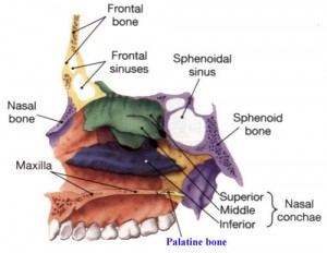
In the human body, this bone is located at the back of nasal cavity – between the pterygoid process of the sphenoid and the maxilla. The Transverse palatine suture joins this bone and the maxilla.
Palatine Bone Ossification
The membrane in the perpendicular plate of this bone begins to appear during the last week of the second month of fetal growth. During this time, the bone gets ossified in this membrane. From this point, the ossification spreads downwards into the pyramidal process, medially into the Palatine Bone’s horizontal plate and upwards into the sphenoidal as well as orbital processes.
Palatine Bone Description
The human Palatine has an L-shaped structure. However, the appearance of this bone may vary slightly in different animal species. It forms a V-shaped structure in various amphibians like the Rough Skinned Newts. The horizontal and vertical elements of this bone make up a 45 degree angle in various species of cats.
Palatine Bone Anatomy
In humans, the Palatum enters into the two fossæ formations: the pterygopalatine fossæ and pterygoid fossæ. It is also connected with the inferior orbital fissure.
The bone comprises of one Horizontal plate, one Perpendicular Plate and 3 outstanding processes known as:
- Pyramidal Process
- Orbital Process
- Sphenoidal Process
The Pyramidal Process of the Palatum is located at the back of the greater palatine foramen and is pointed backwards in a lateral manner from joint of the two human Palatines. The Orbital Process and the Sphenoidal Process is located at the top of the vertical part of the Palatines. A deep notch, known as the sphenopalatine notch, separates these two processes.
The sphenopalatine notch separates the Palatum processes located on the superior border of the bone. The lower surface of the sphenoid converts the sphenopalatine notch into sphenopalatine foramen.
There are several small bumps in the corners of the soft palate where it joins the rear side of the tuberosities. These bumps are known as the hamulii. The hamulii are actually the tips of the little projections from the base of the skull, known as the hamular processes of the palatine bone.
Palatine Bone Articulations
The Palatum is a facial bone and articulates with the following bones:
- Sphenoid bone
- Ethmoid bone
- Maxilla
- Inferior Nasal Concha
- Vomer
- Opposite palatine
Palatine Bone in Other Animals
Bony Fish species have this bone consist of only the Perpendicular Plate. It lies on the inner edge of the maxilla. In fishes, there are generally several teeth arranged on its lower surface, forming another row behind the maxilla. Palatine teeth are often larger, as compared to the maxillary teeth.
Similar patterns could be studied in primitive tetrapods. However, the Palatum merely forms a narrow bar between the maxilla and vomer in existing amphibians such as frogs and salamanders.
In various species of mammals, the lower surface of the Palatine has been found to fold over due to evolution and form the Horizontal Plate, meeting at the middle of the mouth. It can be seen in almost all mammals including canine, feline and equine species. This makes breathing easier during eating by forming the rear side of the hard palate that separates the nasal and oral cavities. Many living reptiles, including the crocodilians, have developed similar Palatines as mammals. In case of birds, the Palatums are separated and form a movable articulation with their craniums.
Palatine Bone Functions
One of the main functions of the Palatine pair is to form a portion of the hard palate or the upper surface (roof) of the mouth. They also make up an important part of the eye sockets and nasal cavity. The Bones of the palatine also give proper shape to the orbits, nasal cavity and oral cavity.
Horizontal Plate of Palatine Bone
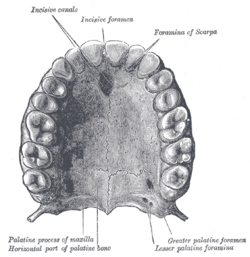
The horizontal plate or horizontal part of the Palatum is a quadrilateral bone, having fur borders and two surfaces. The concave superior surface forms the backside of the nasal cavity floor while the inferior surface forms a portion of the hard palate. Sometimes, there is a transverse ridge marking on the lower surface of the Horizontal Palatum Plate.
The four borders of the horizontal plate are known as:
- Anterior border
- Posterior border
- Lateral border
- Medial border
The medial end of the Horizontal Plate’s posterior border forms an extended process by uniting with the pointed end of the bone opposite to it. It is known as the posterior nasal spine.
Perpendicular Plate of Palatine Bone
The Perpendicular Plate or Vertical Part of the Palatum is oblong and thin, having four borders and two surfaces. One of the two surfaces is known as the Nasal Surface. It has a shallow, broad depression at its lower portion which constitutes an important part of the inferior meatus of the nose. The maxillary surface is irregular and rough, forming an articulation with the nasal surface of the maxilla.
The Perpendicular Plate has the following borders:
- Anterior border
- Posterior border
- Superior border
- Inferior border
Palatine Bone and Cleft Palate
Cleft palate is a congenital condition that occurs if the palatine of a child does not develop properly before birth. Sometimes, the two skull plates that make up the hard palate or the upper surface of the mouth are not properly joined. This condition is known as the cleft palate. In such cases, the soft palate is cleft as well. Most individuals with cleft palate have a cleft lip. This is a fairly rare condition, affecting 1 out of 700 live births in the world. A cleft palate or lip can be treated with surgery.
Palatine Bone Pictures
Take a look at some diagrams of the Palatum that display various parts of the bone.
Picture 1 – Palatine Bone
Picture 2 – Palatine Bone Image
References:
http://en.wikipedia.org/wiki/Palatine_bone
http://www.theodora.com/anatomy/the_palatine_bone.html
http://www.prohealthsys.com/anatomy/grays/osteology/palatine_bone.php


