Carpal Bones are one the most important structures of the human body. Read on to know about the definition, anatomy and functions of these bones.
What are Carpal Bones?
Page Contents
These are eight tiny bones that are found between the five metacarpals and the distal ends of ulna and the radius. These are considerably different in size and are arranged into two rows, each consisting of four bones.

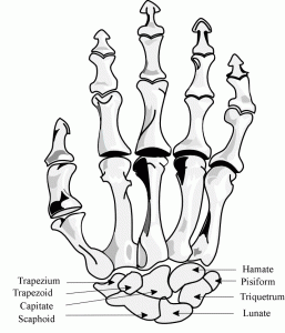
Picture 1 – Carpal Bones
Source – fpnotebook
As the bones are short, they have a compact bone construction that surrounds a porous or cancellous bone center.
The carpal bones come together to form a joint with the long bones of the forearm. It joins proximally with the radius and the ulna and distally with the five metacarpals that cluster together to make the palm.
The bones of the proximal carpal rows are often called as
- Radial bones
- Intermediate bones
- Ulnar bones
- Accessory bones
The bones in the distal row are often regarded as the first, second, third, and fourth carpal bones.
What Does Carpal Bone Mean?
The term Carpal Bone stands for the bones that are located in the Carpus or the wrist. The carpus is a complicated bone joint that allows
- Adduction and abduction in the front level of the upper extremity
- Pronation and supination in the coronal level
- Flexion and extension for hand movements
The eight bones of the Carpus are known as the lunate, scaphoid, capitate, hamate, trapezium,triquetrum, trapezoid, and pisiform.
Anatomy
The proximal carpal bone row lies near the radius and the ulna. It extends from the lateral side or the side of hand that lies farthest from the body to the medial side or the side that lies closest to the midsection of the body. It consists of the bones which are named Lunate, Scaphoid, Pisiform and Triquetrum.
The distal row of carpal bones that lies at the farthest position from the ulna and the radius consists of the Trapezoid, Hamate, Trapezium and Capitate.
The eight carpal bones of the hand, when viewed together, appear as a concave structure from the front. This indicates that they have an inbound curve. It forms a shallow dip or an indent when looked at from the front. When watched from the back, the carpal bones tend to appear as a convex structure, which means that they pop out outward. The indentured carpal bones form the carpal tunnel, under the cover of the flexor Retinaculum.
Function
The anatomical placement of the carpal bones helps them form a joint. Carpal bones of wrist are responsible for rotation and flexibility of the wrist and hand of human beings. Cartilage surrounds four surfaces of the carpal bones. The presence of cartilage allows the bones or the joint to articulate and smoothly move against each another, without causing any discomfort or pain. It is only when this cartilage degenerates that the bones scrape against one another and gives rise to pain that may range from mild to acute.
Injuries
The carpal bones are highly susceptible to injury. This is due to their structure as well as the range of movement in the wrist. Injuries of the carpal bone generally take place while performing sports activities. Injuries may also result from repetitive motion as well as falls. Most injuries take place when the wrist is placed in a flexed position. An injury in the carpal bones gives rise to pain and extreme discomfort.
The carpal bones, ligaments, and tendons are under maximum stress when they are flexed. In case of an injury in the carpal bones X Ray examination is carried out for diagnosis. An X Ray examination helps in accurately depicting broken carpal bones.
Fractures
Usually, a carpal bone fracture does not involve a crack in the skin. This is known as a closed fracture. Carpal fractures may be either unstable or stable. The fragments can displaced out of their usual position or non-displaced in usual alignment. Carpal fractures can arise with joint dislocation or without it. The most common fractures are displaced to a minor degree.
Carpal fractures usually happen when a person suffers a fall and the impact of the plunge is on the outstretched hand. Some other causes involve
- Motor vehicle accidents
- Collisions between participants in sports activities
- A sudden hit to the palm by a golf club or baseball bat
Carpal fractures are quite common injuries in sportspersons, laborers and gymnasts. Stress carpal bone fractures can arise in individuals who are going through repetitive injury such as while using a jackhammer.
The posture of the hand during impact and the intensity of forces exercised on the hand decide which carpal bone is fractured. When baseball bats, sports racquets, or golf clubs hit a static object with excessive force, the curved part of the Hamate bone can suffer a fracture.
Common Carpal Bone Fractures
The carpal bone that is most commonly fractured is the Scaphoid. Scaphoid fractures are the most crippling type of Carpal Bone injuries. They need treatment for a longer period of time. Surgery is commonly used for cure. Scaphoid fractures are more susceptible to complications than other carpal bone fractures.
The second most frequent carpal fracture is the one in the triquetrum. This carpal bone is generally fractured when the person falls on one outstretched hand and the wrist is in ulnar position.
The third most frequent carpal fracture is of the Lunate. Fracture of this bone can result in Kienböck’s disease in which the bone turns granular, fragmented and soft. Kienböck’s disease is supposed to result from repetitive stress.
Approximately 18% of hand fractures happen due to fracture of the Carpal bones. The most commonly fractured bones are the ones in the proximal carpal row. Scaphoid fractures account for about 60 to 70 percent of all carpal bone injuries.
References:
http://emedicine.medscape.com/article/97565-overview
http://www.medterms.com/script/main/art.asp?articlekey=10326
