Mucocele Definition
Page Contents
- 1 Mucocele Definition
- 2 Mucocele Incidence
- 3 Mucocele Location
- 4 Mucocele Symptoms
- 5 Superficial Mucocele
- 6 Appendiceal Mucocele
- 7 Sinus Mucocele
- 8 Mucocele Causes
- 9 Mucocele Diagnosis
- 10 Mucocele Differential Diagnosis
- 11 Mucocele Treatment
- 12 Mucocele Home Treatment
- 13 Mucocele Prognosis
- 14 Mucocele Duration
- 15 Mucocele Complications
- 16 Mucocele Management
- 17 Mucocele Prevention
- 18 Mucocele Pictures
It is a painless cyst that develops on the inner side of the lip due to an accumulation of saliva. The growth is also known by many other names, such as mucous retention cyst, mucus escape reaction and mucus retention phenomenon.
Mucocele Incidence
The condition has a higher prevalence among young people. The cysts also develop commonly in patients who have recently undergone an Endoscopic endonasal surgery.
Mucocele Location
Cysts of this type are most commonly found to arise on the surface of the lower lip. However, they may arise at any spot within the oral cavity. A mucocele cyst that occurs on the gum is referred to as an “Epulis”. A mucous cyst arising on the floor of the mouth is known as a “Ranula”. When located on the inner side of the cheek, they are termed as “Buccal Mucosa”. These may also be found on the anterior ventral tongue. The growths can only rarely be seen to develop over the upper lip.
The buccal mucosa and mucosa of the lower lip are the most common locations for the formation of these hickeys. However, doctors believe that any region that comprises of intraoral salivary glands can be a potential site.
Mucocele Symptoms
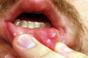
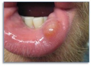
The condition leads to the development of soft, rounded lesions that have a diameter sized anywhere from 1 to 10 mm. It has a smooth surface that comprises of translucent or pearly mucosa. The growths are generally transparent and their color may be light blue, white or the same as that of normal mucosa cysts. When touched, the lesions may remain firm or move under the surface due to finger pressure.
The growths develop quickly and frequently occur on the inner side of the lip. These may persist for a few days or even last for several years. There may be recurrent inflammation of the lesions, with occasional rupturing that may release the contents inside them.
Patients with these hickeys regularly complain about the lesions varying in size from time to time. This has been recognized as an important diagnostic characteristic for this condition. During diagnosis, doctors often look for a cyst that shows variable growth in patients.
These cysts are easily prone to rupture due to trauma. When injured, they release a sticky, straw-colored fluid from within.
Superficial Mucocele
It is a form of mucocele that may develop over the palate, posterior buccal mucosa and retromolar pad. It manifests presents as single or multiple cysts and ruptures into an ulcer. Though these lesions heal after a few days, they are often found to reappear in the same location.
Appendiceal Mucocele
It is a cystic mass of the appendix that develops from abnormal collection of mucus. It is a rare disorder that is found in only 0.07-0.4% cases of all appendectomies. The condition is four times more prevalent in females than males and is usually diagnosed when the patient is around 50 years of age. It is a potentially dangerous tumor that is generally associated with malignancy. However, there are many benign forms of this condition that do not involve any cancerous complications.
This abnormal mass can give rise to extremely discomforting problems, such as:
- Loss of weight
- Nausea
- Vomiting
- Anemia
- Hematochezia
- Hematuria
- Changes in bowel habits
Pseudomyxoma peritonei is the most dreaded complication of both benign and malignant types of mucocele. It is hard to treat this complication, either medically or surgically. It has an unsure prognosis, with approximately 53 – 75% patients showing a 5-year survival rate.
Sinus Mucocele
It is a cyst formed by an accumulation of mucous within the sinus cavity. It is bordered by the mucous-releasing epithelium located within a paranasal sinus. Such cysts usually develop due to the obstruction of a sinus ostium or a septated sinus chamber. This leads to mucus accumulation within the sinus cavity and makes it airless. The obstruction often occurs due to an inflammation. However, it may also develop due to a trauma, surgical manipulation or tumor.
These cysts are generally situated in the Ethmoid, Frontal and in some cases in the Maxillary sinus cavities. These abnormal mucosal growths can give rise to difficult symptoms like,
- Pain
- Nasal obstruction
- Bone deterioration
In severe cases, they may affect the breathing pattern and even lead to blindness. Loss of vision usually results from the development of a large mucocele in the Frontal as well as Ethmoid sinus cavities.
Mucocele Causes
The condition is generally believed to be a result of severe trauma to the salivary ducts. The traumatic rupture of these ducts allows the saliva contained inside them to release into the mucosa. This leads to an accumulation of transparent fluid on the inner surface of the lip. This gives rise to mucus retention cysts over the interior lip surface. The mucosal hickeys may also arise in the buccal mucosa, on the gum or under the tongue.
These mucosal lesions are also indicated to be a result of obstruction of salivary ducts in patients. Both obstruction and rupture of the ducts may occur due to causes like:
Biting or sucking lips
Individuals who have a habit of biting their lip or sucking their inner lip between their teeth are often found to be at risk from this problem.
Piercing
The condition may also be witnessed in people who have recently had their lip or tongue pierced. Constant pressure over the inner lip membranes can serve to block or tear the salivary ducts.
History of an injury
In some cases, patients may report of a remote or recent injury to the face or mouth. During diagnosis, doctors often ask patients about any history of a prior injury to the affected region.
Dentures
An Epulis, or mucous cyst forming over the gum, is usually a result of repeated injury to the affected spot. People who wear dentures are often found to suffer from this type of trauma.
Tongue thrusting
In cases where hickeys develop over the anterior ventral surface of the tongue, trauma as well as tongue thrusting can be the causative factors.
Often, patients having a history of lichen planus or lichenoid drug reaction are found to suffer from disorders involving the oral mucosa. In many cases, however, no cause of Mucocele development can be identified.
Ranulas are often formed as a result of obstruction of the salivary gland ducts, damage to the tissues or trauma.
Mucocele Diagnosis
In most cases, the cyst tends to heal or rupture independently when the impacted region is left undisturbed. Persistent trauma to the impacted area can delay the healing process and possibly even lead to the expansion of the cyst due to the accumulation of extra fluid. If the lesion gives rise to discomforts, it may be necessary for patients to visit a doctor immediately. The diagnosis of the growth can be confirmed by a visual examination of the affected spot.
Mucocele Differential Diagnosis
The differential diagnosis of Mucocele involves distinguishing the disorder from other conditions that can produce similar symptoms. These include:
- Branchial Cleft Cyst
- Dermoid Cyst
- Lipomas
- Lymphangioma
- Oral Hemangiomas
- Oral Lymphangiomas
- Oral Pyogenic Granuloma
- Salivary gland neoplasm
- Varix
- Venous Lakes
Mucocele Treatment
Surgical excision is the best possible option for curing these growths. Excision should be deep enough to reach the underlying gland that causes development of the nodules. Discomforting symptoms can be relieved by cutting open the cyst and allowing drainage. Surgically opening the cysts should best be performed by experienced healthcare providers in a clinic or doctor’s office. This can lessen the possibilities of infection.
If an infection develops in the area, antibiotics are the standard medications used to address the problem.
Surgical removal of these cysts should best be done with the aid of Laser operation and other minimally-invasive procedures. These methods, though expensive, can help drastically reduce the time for recovery.
If symptoms are found to be minimal in young kids and infants, aspiration of the cysts and occasional follow-ups for six months can be useful instead of a surgical operation.
Mucocele Home Treatment
A small or newly detected mucocele can be cured with the aid of home remedies. Salt-water wash is a standard remedy for Mucoceles and quite an effective non-surgical option for tiny cysts of this type. The method involves rinsing the mouth with a cup of lukewarm water mixed with one tablespoon of salt. This should be repeated for four to six times every day for about a week. The solution should be kept in the mouth for 3-5 minutes before spitting it out. The salt helps draw out the fluid trapped beneath the skin surface without causing further damage to the adjoining tissue. If the mucocele prevails, affected individuals should consider visiting a doctor for diagnosis and treatment.
Mucocele Prognosis
The condition generally has a good outcome. This is a benign, common disorder that usually resolves after some time without any external medical assistance. This is especially true in case of mucoceles and ranulas in infants and young children.
Some hickeys turn out to be chronic and need to be removed surgically. Recurrence is common, which can be prevented by excision of the salivary gland adjacent to the affected one. Recurrent mucoceles can lead to permanent scarring of the area on which they develop. Persistent recurrence of these growths can give rise to permanent, firm nodules over the spots where they develop.
Normal mucoceles usually go away with one proper operation. But ranula and epulis cysts require frequent surgical removal to prevent greater discomfort and complication.
Mucocele Duration
The lesions usually persist for three to six weeks before disappearing completely. In rare cases, however, the cysts may take anywhere a few days to a few years to resolve altogether.
Mucocele Complications
In some cases, some forms of mucous cysts may give rise to complications that require medical attention. An affected individual may often rupture or accidentally remove the upper tip of the vesicles by sucking over them. Patients are frequently seen to report of recurrence or a chronic progression of the condition.
Mucocele Management
Patients who have undergone surgical correction for this condition should modify their dietary habits. Diet modifications should depend on the magnitude of surgery. Most patients who undergo oral surgical procedures are usually recommended a diet consisting of liquids or soft foods for the first few days after operation.
More invasive surgeries which involve the excision of a major salivary gland may need to follow a modified diet for a longer duration.
Depending on the magnitude of the operation, patients are asked to stay away from al kinds of strenuous activities for several days or even weeks after surgery. In the post-operative stages, patients are not recommended to use tobacco products until healing has occurred.
Mucocele Prevention
Here are some steps that you may adopt to prevent an occurrence of this condition:
- Avoid sucking your lips whether or not you have mucocele.
- Pick an effective, but mild toothpaste brand that controls tartar. This can help you avoid oral mucocele.
- Chew your food well and eat them slowly. Eating properly can let you avoid causing injuries to the oral region.
Mucocele Pictures
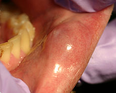
Here are some assistive Mucocele photos that will let you know how the cysts look like. Take a look at these Mucocele images to get an idea about the physical appearance of these cysts.
If you are suspecting a Mucocele development on your oral region, try the salt-water remedy for a few days. If the condition fails to improve over several days, it is best to consult a doctor. Proper diagnosis and care will help you recover from the problem much faster and get back to health sooner than you can imagine.
References:
http://emedicine.medscape.com/article/1076717-overview
http://www.ncemi.org/cse/cse0411.htm
http://www.zhub.com/pathology/listings/39.html
http://dermatology.cdlib.org/1507/case_presentations/sga/lee.html


