What is Femoral Artery?
Page Contents
- 1 What is Femoral Artery?
- 2 Femoral Artery Location
- 3 Femoral Artery Anatomy
- 4 Common Femoral Artery
- 5 Superficial Femoral Artery
- 6 Profunda Femoral Artery
- 7 Femoral Artery Sheath
- 8 Femoral Artery Branches
- 9 Femoral Artery Pulse
- 10 Femoral Artery Diseases
- 11 Femoral Artery Bypass
- 12 Femoral Artery Angioplasty
- 13 Femoral Artery Pain
The Femoral Artery is a term used for a group of few arteries which passes fairly close to the outer surface of the thighs. It begins at the inguinal ligament, called the Femoral Head, and ends just above the knee at p place called the Adductor canal or the hunter’s canal. It divides into smaller branches so as to supply blood to the muscles and to the tissues which lie in the superficial region of the thigh.
Femoral Artery Location
This artery begins immediately behind the inguinal ligament. This place is known as the femoral head. From here, it passes midway between the anterior spine of the ilium and symphysis pubis and continues down the medial and front side of the thigh. At the juncture, where the middle and the lower third of the thigh meet, this artery ends, and here it passes through an opening in the Adductor magnus, and becomes the popliteal artery. The upper third of the artery is contained in the femoral triangle, which is also known as the Scarpa’s triangle, and its middle third is contained in the Hunter’s canal, or the Adductors canal, as it is commonly called.
Femoral Artery Anatomy
The external iliac artery supplies blood to the femoral artery. This artery lies within the femoral triangle, behind the inguinal ligament, usually near the head of the femur bone. This region is known as the inguinal-femoral area. The inguinal ligament borders the trianglre on the superior end, the addustor longus forms the medial border and the lateral border is by the sartorious muscle. The top part of the triangle is made up of skin, fascia lata, cribiform fascia and the subcutaneous tissue. The lower part is composed of iliopsoas muscles, underlying adductor longus and the adductor brevis.
Common Femoral Artery
The proximal section of the femoral artery is known as the Common femoral artery (CFA). It is used as a catheter access artery, as it can be easily felt from the skin. The common femoral artery often comes to use, when the blood pressure is too low, so as to draw arterial blood, as the low blood pressure does not allow the arterial or radial arteries to be located.
This part of the artery is often susceptible to the Peripheral arterial disease. Sometimes the common femoral artery may be blocked through the atherosclerosis. At this stage, we may need to access the Common femoral artery from the opposite side through a Percutaneous intervention. A surgical cut down, called the Endarcetectomy may also help.
Superficial Femoral Artery
The femoral artery leaves the femoral triangle through an apex beneath the sartorious muscle. Here it divides itself into the deep and superficial artery .the superficial branch is called the superficial femoral artery (SFA). This superficial femoral artery connects to the popliteal artery at the opening of the Adductor magnus or the Hunter’s canal at the end of the femur bone.
Profunda Femoral Artery
The profunda femoral artery, also known as the Deep femoral artery, is the posterior branch of the femoral artery. It is the largest branch of the femoral artery in the entire femoral triangle. It arises on the lateral side of the femoral artery, about 3 to 5 cm below the inguinal canal. From here, it travels down the thigh, to the femur, passing between the pectineus and the adductor brevis, and then passes posteriorly behind the adductor longus.
The profunda femoral artery branches into the following:
- Lateral circumflex artery
- Medial circumflex femoral artery
- Perforating arteries
- Terminal branches
Femoral Artery Sheath
The femoral sheath is formed by a downward prolongation, behind the inguinal ligament, transversalis fascia and the iliac fascia. The sheath is in the form of a short tunnel and it is directed upwards. It is also called the crural sheath. This sheath is strengthened by a band called the deep crural arch.
Femoral Artery Branches

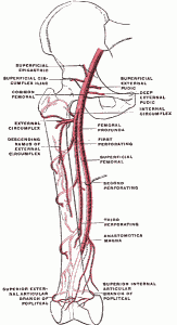
Picture 1 – Femoral Artery and Its Major Branches
Source – wikipedia
The femoral artery has the following branches:
- Superficial Epigastric – This artery arises from the front of the femoral artery, about a cm below the inguinal canal. From here, it travels through the femoral sheath and the fabscia cribrosa, turning upward in front of the inginual ligament, and then ascends between the layers of the superficial fascisa. Its branches are distributed all over the subinguinal lymph glands.
- Superficial Iliac Circumflex Artery – This is the smallest of the cutaneous branches. It arises close to the superficial epigastric artery and runs parallel with the inguinal ligament.
- Superficial External Pudendal Artery – arises medially from the femoral artery, and courses medialwards, across the spermatic cord in males, or the round ligament in females. It is distributed to the integument in the lower part of the abdomen – the penis and scrotum in males and the labia majora in females.
- Deep External Pudendal Artery – this artery is more deeply placed than the superficial pudendal artery. It is covered by the fascia lata, pierces in the middle of the thigh, and is distributed to the intugement of the scrotum and perineum in males, amd to the labius majus in females.
- Muscular Branches – these branches are supplied by the femoral artery t the Vastus medialis, Sartorius and Adductores.
- Highest Genicular
Femoral Artery Pulse
The femoral pulse is located in the inner thigh, at the mid-inguinal point, which lies halfway between the anterior superior iliac and the symphsis. This pulse can be felt at the groin area of the body.
Femoral Artery Diseases
Femoral Artery Occlusion
The Peripheral Arterial Occlusive Disease (POAD) is a disease that results from inflammatory processes or atherosclerotic processes which causes stenosis or narrowing of the lumen. It is also called the Femoral Artery Disease. It may also result due to the formation of the thrombus, as it is often associated with the underlying atherosclerotic disease. An increase in this disease can lead to vessel resistance which in turn leads to a reduction in distal perfusion pressure. The conditions of peripheral arterial occlusive disease are very similar to those found in the coronary artery disease.
POAD occurs mostly in the leg. This is due to the fact that femoral artery is a continuation of the external iliac artery. The deep femoral artery, which is a major branch of the femoral artery, is continues down the leg and becomes the popliteal artery.
The most severe effect of POAD is that it can lead to limb ischemia. In mild conditions, increased resistance to flow may lead to a decrease in flow of blood during limb exercise. This condition is known as decreased active hyperemia. Very acute reductions in perfusion pressure leads to a fall in the vascular resistance and thus maintains the normal blood flow.
Femoral Artery Aneurysm
Aneurysm or widening of the artery normally occurs in the groin as the femoral artery is normally situated in the joint. It is a condition that is demonstrated by the localized, blood-filled balloon like bulging and weakening of the walls of the blood vessels. It can happen in the Aorta or the blood vessels also, apart from occurring in the femoral artery.
Aneurysms are more common in older men than women. The cause of aneurysm is not known. If the aneurysm is very small, then it may not even be seen. However, to some people, the symptoms may appear in the form of a lump, which may even be pulsating. Cramps in the legs while exercising may also cause trouble. The patient may need to seek medical treatment to get cured of this ailment.
Femoral Artery Pseudoaneurysm
Whenever a penetrating injury occurs in the femoral artery, a bubble may be formed on the artery. Since now, there is an opening in the artery; there may be leakage of blood from the artery walls. This feature is called Hematoma, which develops walls around it and then liquefies forming a pulsating bubble, which is known as a Pseudoaneurysm. The most common way by which this feature can occur is during the performance of the cardiac catheterization via the femoral artery.
Femoral Artery Blockage
Sometimes blood clots may occur in the artery due to the atherosclerosis or any other reasons. When this blockage occurs in the leg artery, it is known as the Femoral artery blockage. This blockage may be depicted by the way of severe cramps in the leg and leg aches.
The symptoms of this blocked femoral artery include:
- Cramps in the legs, thighs or calves
- Discoloration of the skin
- Faint pulse in the leg
- Slow healing of wounds
- Impotency in men.
The condition, after diagnosis should be treated at once. A surgery may be required to remove the blockage.
The method of treatment of these artery diseases is:
Femoral Artery Bypass
Femoral popliteal bypass surgery is a surgical method of treating the femoral artery disease. It is the method by which, the upper portion of the leg is surgically opened so as to visualize the femoral artery directly. Pieces of veins taken from legs may be used to perform the bypass surgery. The surgery is performed by attaching one end of the vein above the blockage, and the other below the blockage, thereby rerouting the blood flow around the blockage, through the new graft to reach the muscle. Sometimes, a prosthetic graft may also be used in place of the vein graft.
Femoral Artery Cannulatiuon
This is a standard method of cardiopulmonary bypass (CPB). It ensures a true-lumen cannulation, but it has some disadvantages that include lower extremity ischemia, potential cerebral hypoperfusion and embolization.
Femoral Artery Angioplasty
Another method of surgical treatment of blockage is percutaneous transluminal angioplasty or Femoral artery angioplasty. This is a comparative low invasive procedure to open the blocked femoral artery and to restore the blood flow, without a proper vascular surgery.
During angioplasty, a stent or a catheter tube is inserted in the femoral artery, having a balloon at its tip. This is called vascular stenting or Femoral Artery Stent. A stent is a small wire mesh tube having a balloon kind of structure at its end. When the balloon is inflated, it compresses the fatty tissues in the artery and thereby increases the opening in the artery improving the blood flow.
Femoral Artery Pain
The pain can be caused in the femoral artery due to many reasons. The primary reason is the blockage of the artery. The pain is experienced in the legs or feet, as the blood is unable to flow downwards due to the blockage.
Symptoms
- Sudden or sharp eruption of pain.
- Lower abdomen pain.
- Pains during exercise session.
Causes
- Hernia
- Kidney stones
- Conditions like arthritis, bone dislocations and fractures
- Swollen groin lymph nodes
- Groin pain in women
It is very important to treat the complication that arises in this artery as it is essential for life. Proper treatment should be given and care should be taken the artery is free from any sort of external pressure.
Reference:
http://en.wikipedia.org/wiki/Femoral_artery
http://www.bartleby.com/107/157.html
http://www.innerbody.com/image_cardov/card41-new.html
http://www.vasculardoc.net/content/aneurysms_femoral.html
http://www.ehow.com/about_5539590_femoral-artery-aneurysm-symptoms.html#ixzz1KJssVm9r
http://www.vasculardoc.net/content/aneurysms_femoral.html
http://www.ehow.com/about_5539590_femoral-artery-aneurysm-symptoms.html
http://en.wikipedia.org/wiki/Aneurysm
http://www.ehow.com/about_5482226_femoral-artery-pain.html
http://www.dartmouth.edu/~anatomy/Lowerextremity/vessels/tutorial/femoral.htm


Aloha,
I had Inguinal Hernia surgery on 7-2-11 in Elkhorn, WI with Atrium Pro-Loop Mesh and Mesh Plug. I have been in pain ever since. Is there anyother’s that you have heard that have had this also? Please let me know. Thank you!
I had that surgery also and it took 3 weeks for pain to subside enough for me to go back to work. Yes very painful I feel for you.
Since December 2014 I have incredible pain in my groin, the back of my thigh and the front. It has caused weakness. The pain and weakness have been so bad that I cannot drive and I can not find a comfortable position to sit or sleep. It hurts MUCH worse sitting than walking. The bones are fine. (Had an X-Ray done.)
Since December 2014 I have incredible pain in my groin, the back of my thigh and the front. It has caused weakness. The pain and weakness have been so bad that I cannot drive and I can not find a comfortable position to sit or sleep. It hurts MUCH worse sitting than walking. The bones are fine. (Had an X-Ray done.